Breast Biopsy
A breast biopsy may be needed to remove a small portion of tissue or fluid for further testing. Our breast care team conducts several types of biopsies:
Core needle biopsy – A small needle is guided into the lump or suspicious area. Several small areas of tissue, called cores, are removed for testing.
Stereotactic core biopsy – A 3D image of the breast is created with your mammogram images. The radiologist uses the 3D image to guide the needle to the exact site of the lump or suspicious area.
Vacuum-assisted core biopsy – A suction device is used to pull tissue and fluid gently into a hollow needle, to increase the amount of cells removed for testing.
Fine needle aspiration – A very thin needle is inserted into the lump or suspicious area. A sample of tissue or fluid is removed for testing.
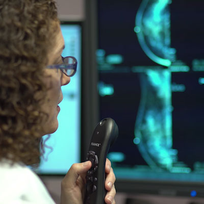
Image-guided biopsy – Radiologists sometimes use an imaging procedure — such as ultrasound or magnetic resonance imaging (MRI) – to help guide the needle to the exact area for needle biopsy.
Needle localization for surgical biopsy – In some cases the lump may be hard for your doctor to reach. A needle with a thin wire is inserted into the lump using imaging. The surgeon then follows this wire to find the lump and remove a tissue sample for testing.